

Porcine Skin Be Prepared and Used in In Vitro Skin Permeation Studies

Introduction
Transdermal administration is an attractive route of administration because it is easy to administer and carries a relatively low risk of systemic effects, which enhances the safety of treating localized conditions such as dermatitis. However, if systemic effects are required, formulations capable of transdermal delivery of therapeutic agents can also be used. Because the skin acts as a semipermeable barrier, various factors may influence the permeability of drugs applied to the skin, including ethnicity, skin hydration, age, and the presence of absorption enhancers in the formulation. The physicochemical properties of drugs and formulations also influence drug permeability through the skin, as well as the pathways taken by the drug during the permeation process. Drugs can follow three distinct pathways: intercellular (between keratinocytes), transcellular (through the keratinocyte membrane), and via skin appendages (through hair follicles). The Organization for Economic Cooperation and Development (OECD) and the U.S. Food and Drug Administration (FDA) recommend the use of the Franz diffusion cell system to simulate drug diffusion through the skin. Human skin is the membrane of choice for such studies and can be obtained surgically. However, given the limited availability of human skin and
restrictions on access, membranes from other animal sources are commonly used. Among these, porcine ear skin is the most suitable membrane; numerous studies indicate that the use of porcine ear skin is the gold standard for simulating human skin in various in vitro permeation studies. Because porcine ear skin is similar to human skin in terms of stratum corneum (SC) thickness, epidermal thickness, hair follicle structure, vascular anatomy, collagen fiber arrangement in the dermis, and ceramide (sphingolipid) content in the SC, the experimental results obtained are comparable to those from human skin. The acquisition and utilization of biological membranes are critical aspects of in vitro permeation studies. Some literature has emphasized key points that should be standardized in such studies to ensure high-quality results, as well as important considerations when using porcine ear skin (such as the distribution of esterase activity and techniques for simulating damaged skin). However, to the best of our knowledge, no previous publication has aimed to standardize the process of selecting, preparing, and using porcine ear skin as a biological membrane for in vitro skin permeation studies. Therefore, in this article, we refer to the paper by R. Silva (2022) and provide optimizations to the process of obtaining and processing porcine ear membranes (Basic Protocol 1) as well as details regarding their application in in vitro skin permeation studies of drugs and other molecules (Basic Protocol 2). Because these steps are crucial for achieving high-quality results, they help minimize variability in the evaluation process.
Get Free Quote
| Test apparatus | Franz Diffusion Cell System |
| Biomembrane | Porcine ear skin, full-thickness (~1.2 ± 0.2 mm) |
| Standard replicates | 5–6 diffusion cells per formulation |
| Typical protocol time | ~2 days for membrane preparation, plus an 8–24 h permeation run |

1.Preparation of Porcine Ear Skin Membranes (Basic Protocol 1) 1.1 Material

Figure 1: Step-by-step evaluation and cleaning of a pig’s ear. (A) The skin of the pig’s ear shows no damage. (B) The skin is washed with running water. (C) Hair is removed from the skin using a surgical hair 1.2 Collection and Preparation of Porcine ear skin ① At the slaughterhouse, select porcine ears that show no physical or pathological damage. Once the porcine ears are removed, ensure they are immediately placed in a foam cooler containing crushed ice. This is a critical step to minimize deterioration and ensure the ears remain chilled until the time of peeling. The slaughterhouse should be located as close to the laboratory as possible, and the time between ear collection and membrane extraction should be standardized to avoid variations in the study protocol. In our study, we standardized the maximum time between extracting the porcine ear membranes and using them for extraction-permeation studies to 6 hours. ② In the laboratory, before handling the ears, clean the work area with neutral soap and water, and disinfect it with 70% (w/w) ethanol. This cleaning procedure must be repeated at the end of the process. ③ After cleaning the work area, remove the ears from the ice-filled container and visually inspect them to ensure there is no physical or pathological damage (Figure 1A). If the skin quality of the ear is sufficient, rinse it under running water (Fig. 1B) and carefully pat it dry with absorbent paper towels. Then, using surgical scissors, carefully trim the outer areas and edges of the ear (Fig. 1C). All these steps must be performed with care to avoid damaging the ear’s skin during processing. Additionally, each ear should be processed individually to maintain its refrigerated state until the exact moment of processing. Figure 2 illustrates the various layers of the skin.

④ Secure the cleaned and shaved ear to a cork board, and use dissecting scissors to cut away the muscle closest to the base of the ear, as shown in Figure 3A. To facilitate this step, it is best to make an incision along the edge of the ear. Since muscle tissue and cartilage are denser than skin, they are easily identifiable. ⑤ Using dissection forceps (straight, serrated) and a scalpel, remove the skin from the rest of the ear structure (Figure 3B). The scalpel blade should be directed toward the muscle, not toward the skin, to avoid any damage to the skin. ⑥ Once the skin of the ear has been completely excised, secure it to the cork board with the epidermis facing down. Using tissue forceps (1×2-toothed) and a scalpel, the skin should be stretched taut to facilitate the removal of subcutaneous fat tissue (Figure 3C). A light box can be used to better visualize the subcutaneous tissue, making the removal more efficient and minimizing the risk of tissue damage. In this step, it is extremely important to hold the scalpel blade at an angle of approximately 45° to the skin to avoid penetrating the tissue, which could affect the results of subsequent permeability studies.

Figure 3: The process of skin excision from a pig’s ear. (A) Make an incision at the base of the ear to begin separating the skin from the ear’s muscles and cartilage. The process of separating the skin from the ear’s muscles and cartilage begins. (B) The skin is removed from the pig’s ear using dissecting forceps (straight, serrated) and a scalpel handle fitted with a scalpel blade (for dissecting subcutaneous and muscle tissue). (C) Subcutaneous tissue is excised from the skin using tissue forceps (1×2 teeth) and a scalpel handle fitted with a scalpel blade. ⑦ Once the subcutaneous tissue has been removed, the skin can be used immediately for experiments or placed in a sealed plastic bag using a manual vacuum pump. During packaging, ensure that the skin is not bent inside the sealed plastic bag. Once the skin has been properly placed in the plastic bag, it should be stored immediately in a freezer at -20 °C and kept there until the day of the experiment (Basic Protocol 2). Important Note: The timing and method of biofilm packaging are factors that may influence research results. Therefore, each laboratory should evaluate these aspects based on the characteristics of the drug being studied and the enzyme systems involved in its metabolism (if relevant). In our laboratory, we have determined that biofilms must be placed in sealed plastic bags and stored at -20 °C (freezer) for up to 3 months, in accordance with recommendations from guidelines such as the “European Cosmetics: Transdermal Absorption/Permeation Guidelines” and the “Basic Criteria for the In Vitro Assessment of Skin Absorption of Cosmetic Ingredients by the Scientific Committee on Consumer Safety (SCCS).”

2. Preparation of a membrane from porcine ear skin and its use in in vitro skin permeation studies (Basic Protocol 2) 2.1 Materials

Figure 4: Harvesting the skin membrane from a pig’s ear. (A) Assembly diagram of the Franz diffusion chamber. (B) A membrane ring on the skin marked by a biopsy punch. 2.2 Preparation of Membranes for In Vitro Skin Permeation Studies ① If the biological membrane is frozen (-20 °C) after being removed from a pig’s ear, remove it from the freezer 1 hour before the start of the study and allow it to stand at room temperature. Record the room temperature used and maintain it at a constant level throughout the study to minimize the risk of variations caused by temperature changes. Skin surface temperature can be altered by ambient temperature and may affect the skin penetration of drugs applied to the skin, as confirmed in several reports. ② Cut the skin into appropriate sizes to serve as biological membranes in the Franz diffusion experiment; it is best to use a skin biopsy punch. The size of the membrane cut by the punch should match the diameter of the Franz diffusion cell (Figure 4A). The biopsy punch should be placed on the skin at a 90° angle, and sufficient pressure should be applied to cut it, forming a symmetrical membrane (Figure 4B). After cutting the membrane, it is helpful to measure its thickness with a caliper. In our study, the membrane thickness was 1.2 ± 0.2 mm. This range is consistent with values found in the literature (1.0, 1.4, and 1.4–1.5 mm). ③ Place the membrane in the Franz diffusion chamber with the epidermis facing upward, and fill the receptor compartment with PBS (pH 7.4). Allow the membrane to stand for 1 hour to hydrate. ④ After the skin hydration period (1 hour), use a tubing connected to a syringe to completely remove the PBS from the receptor compartment through the sampling port of the Franz diffusion cell (Figure 4). Fill the receptor compartment with the receptor fluid to be used in the permeation study. It is very important to select a receptor fluid that can maintain sink conditions in the receptor compartment. ⑤ To prevent the receptor fluid from evaporating from the receptor chamber, we recommend covering the outlet (sampling port; Figure 4A) of the Franz diffusion cell with a high-viscosity film. When introducing any liquid (PBS or receptor fluid) into the receptor chamber, ensure that there are no air bubbles inside the chamber. This is crucial for maintaining contact between the entire surface of the biofilm and the receptor fluid, thereby preventing variations in drug permeation across the Franz diffusion cell. Bubbles should be removed from the sampling port (Fig. 4A) by tilting the cell. If possible, the receptor fluid should be degassed (e.g., by ultrasonication) before being added to the receptor chamber.

Figure 5. Post-extraction handling of the membrane from the Franz diffusion chamber. (A) The membrane is secured to a cork board, and the cotton swab on the membrane is properly handled to remove excess formulation. (B) The epidermis is removed from the biofilm using a scalpel. The arrows indicate the removed epidermis. 2.3 Application of Formulations and Determination of Drug Concentrations in the receptor fluid and Biofilm Layer ① Use an automatic pipette to apply the sample to the surface of the porcine skin membrane in the top (donor) chamber (at a concentration of 30 mg/cm²) . In the studies cited here, we used three different topical formulations: an emulsion, a gel-solid lipid nanoparticle, and a gel-nanostructured lipid carrier, loaded with fluconazole (1% w/w), adapalene (0.1% w/w), and tretinoin (0.05% w/w), respectively. The formulations were then applied to each pig skin membrane using cotton swabs to form a uniform layer on the epidermis, taking care to prevent the formulations from seeping beneath the donor chamber to ensure they did not come into contact with the receptor fluid. (The amount of formulation retained on the cotton swabs used to apply the formulation to the surface of the skin membrane must be determined: prior to the start of the study, each swab used to apply the formulation to the skin membrane must be placed in a beaker, weighed on an analytical balance, and its weight recorded (one swab per Franz diffusion cell). After applying the formulation to the membrane, each cotton swab must be returned to its respective beaker, weighed again on an analytical balance, and the new weight recorded accordingly. The amount of formulation retained on the glass cotton swab corresponds to the difference between the final weight and the initial weight of the swab. In our study, the total application volume retention rate of the formulation on the cotton swabs was typically observed to be 11%–15%.) ② Allow the topical formulation to come into contact with the biofilm for a period of time, then collect samples of the receptor fluid at the specified time for quantification;The sampling protocol and duration of the study should be determined based on the characteristics of the formulation. If, under normal conditions of use, the preparation remains on the skin for approximately 8 hours, its permeability should be evaluated for at least 8 hours.In our study, drug permeability was evaluated over an 8-hour period, with samples collected from the receptor compartment at 2, 4, 6, and 8 hours. At these predetermined time intervals, a specified volume of receptor fluid must be collected using a syringe with a tubing (it is recommended to use an automated sampling device). The collected receptor fluid must be transferred to a suitable container and sent for drug quantification. The receptor chamber must then be immediately replenished with an equal volume of clean, preheated receptor fluid. During this step, it is essential to ensure that no air bubbles are present on the membrane surface, as these may affect the reproducibility of the results. If air bubbles are present, tilt the Franz diffusion cell to remove them through the sampling port (Figure 4A). In our study of fluconazole, the drug could be quantified in the collected samples. However, for retinoids, the drug was not detected in samples collected at different time intervals. ③ At the end of the study, remove all receptor fluids, and then remove excess formulation from the membrane surface using an appropriate method, such as one of the methods illustrated below. a.In our study of emulsions, excess formulation was removed by washing the membrane surface twice with 500 microliters of an aqueous solution containing 1% (w/v) polyoxyethylene-20 oil-based ether (a nonionic surfactant), applied without removing the donor chamber from the Franz diffusion cell. Subsequently, the membrane was carefully removed from the Franz diffusion cell holder and secured with a needle, dermis-side down, on a cork board covered with plastic film (Figure 5A). The membrane was then dried with a cotton swab. b. In studies of tretinoin-based drugs, a cotton swab soaked in PBS (pH 7.4) was gently swabbed across the skin to remove excess formulation (Figure 5A). In these studies, using three consecutive cotton swabs soaked in PBS (pH 7.4), followed by a dry cotton swab, was sufficient to remove excess formulation without removing the drug from the stratum corneum. We recommend verifying the number of times this step must be repeated based on the characteristics of the formulation. The swabs used to remove excess formulation must be placed in a collection vial for drug quantification. (Solutions and swabs used to remove excess formulation from the membrane surface and donor chamber; see the previous section for details): In our fluconazole study, the contents of each Franz diffusion cell were transferred to a capped vial containing 50 mL of mobile phase (45:55 [v/v] methanol/0.025 M phosphate-buffered saline [PBS], pH 7.0), as fluconazole has an affinity for this solution and can be effectively extracted. Subsequently, the vials (one per Franz diffusion cell) were subjected to 2 minutes of ultrasonication and manual stirring to ensure complete dispersion of the formulation and solubilization of the drug. The solution in each vial was then filtered through a 0.45 μm pore size filter, and the drug was quantified by HPLC. A suitable analytical method must be selected in advance; in our study, approximately 70% of the drug was retained at the end of the study (8 hours). ④ To quantify the amount of drug retained in the skin layers, the skin was stretched over a cork board, and the epidermis was separated from the dermis using a scalpel, with the blade moved in short strokes parallel to the skin. The epidermis is the thinner layer and is easily exposed by the scalpel (Figure 5B). Transfer the removed epidermis and the scalpel blade used to remove it to a vial containing an appropriate solvent capable of extracting and quantifying the drug. (To quantify the drug retained in the epidermis, the scalpel used to remove the epidermis and the epidermal fragments must be placed in a stoppered vial (Vial 1), with one vial per Franz diffusion cell. To quantify the drug in the dermis, the scalpel used to puncture the dermis and the dermal fragments must be added to a separate stoppered vial (Vial 2), with one vial per Franz diffusion cell. In our study of fluconazole, 2 mL and 4 mL of mobile phase were added to Bottle 1 and Bottle 2, respectively, and the samples were allowed to stand for 12 hours. The epidermis (Bottle 1) was then homogenized for 2 minutes at 24,000 rpm using a homogenizer, and the dermis (Bottle 2) was mixed for 1 minute using a vortex mixer. Next, the samples were centrifuged at 1,000 × g for 5 minutes, and 1 mL of the supernatant was aliquoted into a conical glass tube containing 2 mL of 1 M HCl. The tube was vortexed, and 4 mL of ethyl acetate was added. The mixture was homogenized and centrifuged at 1,000 × g for 3 minutes, after which the aqueous phase was transferred to a clean conical tube. Next, add 1 mL of 1 M NaOH and 4 mL of ethyl acetate, vortex the mixture for 2 minutes, and centrifuge at 1000 g for 3 minutes. Transfer the organic phase to a glass bottle and evaporate to dryness under a nitrogen stream at 45°C. Dissolve the residue in 2 mL of mobile phase, filter the solution through a 0.45-μm pore-size filter, and analyze it using the selected HPLC method. In the studies of adapalene and tretinoin, all vials were made of amber glass (to protect retinoids from light) and contained 2 mL (Vial 1) or 1 mL (Vial 2) of methanol (to allow for complete dissolution). The vials were vortexed for 2 minutes, allowed to stand for 1 hour, and then vortexed again for 2 minutes. The resulting solution was filtered through a 0.45-micrometer pore-size filter and analyzed using the selected HPLC method. In our study of fluconazole, 10% and 5%–11% of the applied dose were quantified in the epidermis and dermis, respectively, for the different emulsions evaluated. In the study using tretinoin, 0.6% and 20.5% of the applied dose were quantified in the epidermis and dermis, respectively; in the study using adapalene, 1.2% and 16.4% of the applied dose were quantified in the epidermis and dermis, respectively. The dose applied to each membrane was calculated by subtracting the amount of formulation retained on the glass swab from the average dose (determined during the dose-standardization process).) ⑤ After removing the epidermis, the dermis is cut into small pieces and stored in flasks containing an appropriate solvent for drug extraction and quantification. In our study, prior to drug quantification by HPLC, we standardized the samples by storing them at 4°C for up to 24 hours. However, it is important to use a drug quantification method that can visualize signs of drug degradation should it occur. In our study, drug quantification was performed using HPLC, which allows for the identification of degradation peaks should degradation occur. Drugs retained in the epidermis and dermis can be extracted using a homogenizer and/or vortexing, depending on the drug’s interaction with the tissue. Therefore, it is crucial to standardize this step based on the drug’s characteristics. To determine the amount of formulation applied to the skin, the mass retained in the glass swab (determined by the difference in weight of the swab before and after application) is subtracted from the mass dispensed from the pipette (as determined during pipette calibration).

2.4 Data Analysis (Following Quantification of Drug Concentrations in the Epidermis, Dermis, and Receptor Fluid) ① First, calculate the skin area to which the formulation was applied. This requires knowing the radius of the Franz diffusion cell used; the area is calculated using the following formula (the formula for the area of a circle): ②After quantifying the tretinoin (TRE) content in the skin layers, the TRE content for each application area can be calculated. In our study, we found an average of 32.15 μg of TRE in the epidermis and 0.94 μg of TRE in the dermis. The amount of TRE in each skin area was calculated using the following formula:
3. Discussion 3.1 Background Information Following the application of topical formulations, drug penetration into the skin is one of the factors that must be evaluated during the development of such formulations, as it affects their efficacy and safety. Currently, in vitro assessments of drug diffusion through the skin are conducted using biological membranes and Franz diffusion cells. Among existing animal membranes, biological membranes derived from porcine ear skin are considered to be the most similar to human skin. However, the process of obtaining these membranes must be standardized, and permeation studies must be conducted to minimize errors and obtain reliable results with low variability. Therefore, in this article, we aim to detail these steps and highlight the key points that must be carefully examined and standardized. To obtain porcine ears with healthy skin (free of physiological or pathological damage) while avoiding the introduction of variables in permeation studies, it is important to select an authorized slaughterhouse. Since animals living in confined environments are prone to injury, the quality of porcine ears must be strictly controlled. Therefore, it is necessary to select a sufficient number of visually intact ears and store them on ice immediately after removal from the pig to minimize issues related to deterioration. When using this standardized procedure, 5 or 6 diffusion chambers per assay are generally sufficient. To minimize the risk of perforation during membrane extraction and preparation, training for researchers is essential. After removing the subcutaneous tissue, the biofilm can undergo Transepidermal Water Loss (TEWL) assessment as a strategy to verify tissue integrity. Additionally, establishing a standardized TEWL measurement protocol is crucial. In our study, we found that the TEWL of the membrane after subcutaneous tissue removal was 3–5 g/m²·hr.

3.2 Key Parameters and Troubleshooting To obtain sufficient membrane area and thereby achieve stable and reproducible permeation results, several important parameters must be considered. Table 1 discusses the main key points and strategies for addressing issues.
3.3 Statistical Analysis Graphical and statistical analyses can be performed using statistical software such as GraphPad Prism. First, the assumptions of normality and homoscedasticity must be checked. If these assumptions are met, parametric tests may be used; if not, nonparametric tests should be used to evaluate the groups. Generally, a p-value of < 0.05 is considered statistically significant. Experimental values can be reported as mean ± standard deviation. Regarding the number of replicates, generally, if the protocol is standardized, 5 or 6 diffusion chambers per formulation are sufficient.

Figure 6: An overview of the steps involved in preparing and processing porcine ear skin to obtain a biofilm for use in in vitro permeation 3.4 Interpretation of Results Figure 6 outlines the steps required to prepare and process porcine ear skin to obtain biofilms and to use the biofilms for in vitro permeation studies. Figures 7–9 and Table 2 illustrate examples of in vitro permeation studies we conducted using biofilms obtained from porcine ear skin.

Figure 7 In vitro skin penetration of fluconazole in an emulsion through porcine ear skin. Formulations B and D are emulsions containing 1% w/w fluconazole. Each data point represents the mean (n = 3) ± SD. Reprinted from Ayub et al. (2007), “Topical Administration of Fluconazole: In Vitro Skin Permeation and Absorption,” Drug Development and Industrial Pharmacy. Copyright (2007), used with permission from Taylor & Francis Ltd. (www.tandfonline.com). a: Apply 62 ± 2 mg of emulsion (1% fluconazole) to the skin and spread it evenly using a glass swab; weigh the glass swab before and after application to determine the applied dose (54 ± 2 mg). Reprinted from Ayub et al. (2007), “Topical Administration of Fluconazole: In Vitro Skin Permeation and Absorption Using an Emulsion as a Dosage Form,” Drug Development and Figure 6: An overview of the steps involved in preparing and processing porcine ear skin to obtain a biofilm for use in in vitro permeation Figure 7 In vitro skin penetration of fluconazole in an emulsion through porcine ear skin. Formulations B and D are emulsions containing 1% w/w fluconazole. Each data point represents the mean (n = 3) ± SD. Reprinted from Ayub et al. (2007), “Topical Administration of Fluconazole: In Vitro Skin Permeation and Absorption,” Drug Development and Industrial Pharmacy. Copyright (2007), used with permission from Taylor & Francis Ltd. (www.tandfonline.com). Industrial Pharmacy. Copyright (2007), licensed by Taylor & Francis Ltd. (www.tandfonline.com). Figure 7 and Table 2 illustrate the in vitro permeation of an emulsion (1% [w/w] fluconazole) through porcine ear skin using a Franz diffusion cell (membrane surface area of 1.77 cm², receptor volume of 6.7 mL). In this study, sink conditions were achieved in the receptor chamber using phosphate-buffered saline (PBS) with a pH of 7.4. Due to the characteristics of the formulation being evaluated (i.e., an emulsion containing more than 20% oil phase), we washed the treated surface with 500 μL of a nonionic surfactant (1% (w/v) polyoxyethylene-20 oil-based ether in distilled water) and 500 μL of water to remove excess formulation from the skin. cotton swabs were used to remove any remaining emulsion residue. In this study, the wash solution, pipette tips, and cotton swabs were transferred to a vial containing 50 mL of mobile phase for HPLC analysis of their total fluconazole content. To extract fluconazole from the epidermis and dermis, these fragments were placed in glass vials containing 2 and 4 mL, respectively, of a 45:55 (v/v) mixture of methanol and 0.025 M phosphate-buffered saline [PBS], pH 7.0. The mixtures were allowed to stand for 12 hours, after which the epidermis was crushed using an Ultra-Turrax T 25. Recovery results showed that 92 ± 3.6% of the applied fluconazole was recovered (from the epidermis, dermis, receptor fluid, and materials in contact with the formulation, such as cotton swabs). Table 2 shows that the amount of fluconazole permeating through porcine ear skin ranged from 8% to 9% of the applied dose. The two emulsions retained similar amounts of fluconazole in the epidermis (∼ 10%), but the amounts retained in the dermis ranged from 5% to 11%. Figure 7 shows similar permeation patterns for fluconazole loaded in emulsions B and D.

Figure 8. Intradermal distribution of solid lipid nanoparticles loaded with adapalene and a commercially available adapalene gel. Data are presented as mean ± standard deviation (n = 5). Different letters indicate samples with significant differences (p < 0.05) as determined by Tukey’s post hoc test. Reprinted from Ayub et al. (2007), “Topical Administration of Fluconazole: In Vitro Skin Permeation and Absorption Using an Emulsion as a Dosage Form,” Drug Development and Industrial Pharmacy. Copyright (2007), licensed by Taylor & Francis Ltd. (www.tandfonline.com).

Figure 9. Results showing the amount of tretinoin retained in the epidermis and dermis of porcine ear skin 8 hours after in vitro permeation studies (μg/cm²), expressed as mean ± standard deviation (n = 5). *, statistically significant difference (p < 0.0001); #, below the detection limit of this method. Reproduced from *Nanomedicine* (2021) 16(16), 1391–1409, with permission from Future Medicine Co., Ltd. In both studies shown in Figures 8 and 9, porcine ear skin and a static 20-mm Franz diffusion cell were used, with a diffusion area of 3.14 square centimeters and a receptor volume of 10 milliliters. The receptor fluid was a 90:10 (v/v) mixture of PBS (pH 7.4) and ethanol containing 2% (w/v) polysorbate 60 to ensure sink conditions for the two drugs under study, adapalene and tretinoin. The skin penetration of adapalene (AD) was compared between a formulation containing solid lipid nanoparticles (SLNSA-AD) and a commercially available gel (MKT-AD). Similarly, the skin penetration of tretinoin (TRE) was compared between a formulation containing a nanostructured lipid carrier (NLC-TRE) and a commercially available cream (MKT-TRE). In both studies, 100 μL of each formulation was applied to porcine ear skin, and the skin and receptor fluid were evaluated after 8 hours of exposure. In neither study was the drug detected in the receptor fluid, indicating that no drug permeated through the porcine ear skin. However, in both studies, the drugs permeated into the epidermal and dermal layers of the skin. In the epidermal (16.36 ± 1.79 μg/cm²) and dermal (1.17 ± 0.12 μg/cm²) layers of porcine ear skin, the SLNSA-AD gel retained more AD than the MKT formulation (4.08 ± 0.22 μg/cm² and 0.16 ± 0.03 μg/cm², respectively; Figure 8). In the study of TRE, the nanoparticle formulation also enhanced drug penetration into the dermis and epidermis. Compared with the MKT formulation (0.50 ± 0.07 μg/cm²), the NLC-TRE gel increased the amount of TRE retained in the epidermal layer of the porcine ear by a factor of 20 (10.24 ± 1.06 μg/cm²); TRE reached the dermis only when delivered via the NLC formulation (0.30 ± 0.04 μg/cm²). In both studies, excess formulation on the skin surface could be easily removed using a cotton swab soaked in PBS, followed by drying with a dry cotton swab.
4. Time Recommendations 4.1 Basic Protocol 1 Obtaining porcine ear membranes takes 2 days. On the first day, it is important to prepare the necessary materials (2–3 hours), as thorough preparation of the entire experimental process is crucial for minimizing the time spent handling the porcine ears and ensuring high-quality ear membranes. On the second day, porcine ears should be collected in the morning. The process of harvesting the porcine ear skin from each ear—including disinfection, separation of subcutaneous and muscle tissues, removal of subcutaneous tissue, sealing the porcine ear skin in airtight plastic bags using a manual vacuum pump, and storage at −20 °C—takes 1 to 2 hours.

4.2 Basic Protocol 2 To prepare the membranes for in vitro studies, remove them from the freezer 1 hour in advance. Cut them to the appropriate size; this takes approximately 5 minutes per membrane. Hydration of the membranes takes approximately 1 hour. Remove the buffer from the receptor chamber (Figure 4A) and replace it with receptor fluid; this takes approximately 5 minutes per well. Apply the formulation and spread it evenly over the membrane; this takes approximately 5 minutes per well. (The use of automated sampling equipment is recommended.) The duration of contact between the formulation and the biofilm, as well as the intervals for collecting receptor fluid for quantification, should be determined based on the characteristics of the drug and formulation. However, typically, the evaluation period for drug permeation ranges from 8 to 24 hours to simulate conditions of use. During this period, at least four samples are usually collected to assess drug permeation. To quantify the amount of drug retained in the skin layers, the epidermis and dermis should be separated using a scalpel; this takes 20 to 30 minutes per membrane. To extract the drug that has permeated into the membrane, it is necessary to evaluate the specific characteristics of each formulation and drug, as well as the extraction process.

With the RT8 Series Automatic Sampling Transdermal Diffusion System, users can configure the system to perform automatic sampling, or manually collect samples using a pipette.
Automatic Sample Collection References: Silva IR, Lima FA, Reis ECO, Ferreira LAM, Goulart GAC. Stepwise Protocols for Preparation and Use of Porcine Ear Skin for in Vitro Skin Permeation Studies Using Franz Diffusion Cells. Curr Protoc. 2022 Mar;2(3):e391. doi: 10.1002/cpz1.391. PMID: 35290730.
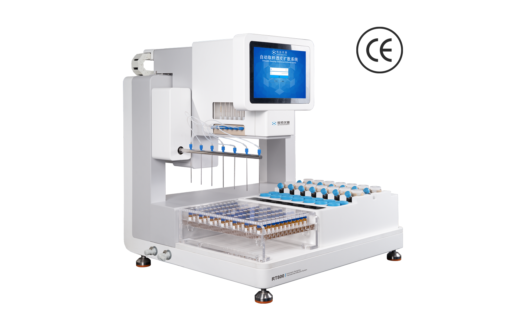
RT800 Automated Transdermal Diffusion System
- Complies with regulation USP<1724>
- 7 channels synchronized automatic sampling
- Meet audit trail requirements
- Shorter pipeline design effectively reduces sample residue
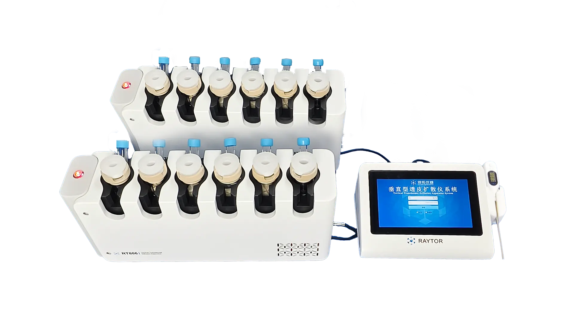
RT806 Vertical Franz Diffusion Cell Tester
- Entry-level transdermal diffusion system
- Compliant with USP<1724> regulations
- Extremely simple structure
- Convenient manual operation

RT814 Automated Transdermal Diffusion System
- Flexible sampling methods
- Real-time monitoring and recording
- Integrated diffusion cell system
- Independent temperature control for all cells