

In Vitro Release Testing IVRT for Ophthalmic Ointments, Gels and Suspensions
2026-07-22


In Vitro Release Testing IVRT for ophthalmic products measures the rate and extent at which an active pharmaceutical ingredient is released from an ointment, gel, or suspension into a receptor medium under controlled laboratory conditions. The method can support formulation development, batch comparison, process evaluation, stability studies, and investigation of critical quality attributes.
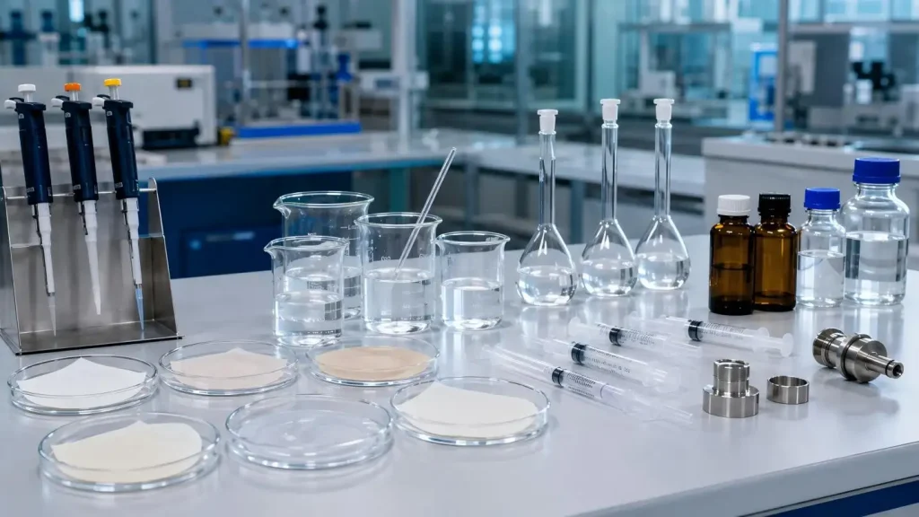
Ophthalmic products are difficult to evaluate because viscosity, microstructure, particle characteristics, and drug solubility can all influence the measured release profile. Reliable In Vitro Release Testing IVRT therefore depends on an appropriate diffusion-cell configuration and strict control of sample loading, membrane selection, receptor conditions, temperature, agitation, and sampling.
What Does Ophthalmic IVRT Measure?
Ophthalmic IVRT evaluates drug release from the dosage-form matrix. In a typical test, the formulation is placed in the donor compartment of a Franz diffusion cell and separated from a temperature-controlled receptor medium by a selected synthetic membrane.
Samples are collected periodically and the total cumulative drug release for that area as well as the release rate are assessed.
In Vitro Release Testing IVRT is particularly useful for evaluating:
•Formulation composition and excipient changes
•API particle size, polymorphic form, and solubility
•Batch-to-batch manufacturing consistency
•Mixing, homogenization, and filling processes
•Product changes during stability studies
•The discriminatory capability of a proposed quality-control method
IVRT does not directly measure corneal permeation, ocular tissue exposure, or clinical efficacy. These endpoints may require complementary permeation, pharmacokinetic, or clinical studies.
Why Are Ophthalmic Formulations Difficult to Test?
Ointments, gels, and suspensions do not release drugs through the same mechanism. Applying one standard method to all three dosage forms may generate poorly discriminatory or highly variable results.
| Dosage Form | Primary Release Process | Main IVRT Challenges | Critical Controls |
| Ophthalmic ointment | Drug partitions from an oily or semisolid vehicle | High viscosity, difficult loading, nonuniform distribution | Sample thickness, temperature, membrane contact |
| Ophthalmic gel | Drug diffuses through a polymer network | Swelling, rheological changes, air entrapment | Gel structure, loading pressure, agitation |
| Ophthalmic suspension | Solid particles dissolve before dissolved drug diffuses | Sedimentation, agglomeration, particle-size effects | Homogeneity, particle retention, sampling strategy |
Ophthalmic suspensions are especially complex because the apparent release rate can be controlled by both particle dissolution and molecular diffusion. Important variables include:
•API particle-size distribution
•Crystal form and surface area
•Dissolved-to-undissolved drug ratio
•Sedimentation and particle aggregation
•Surfactant concentration
•Receptor-medium solubilization capacity
•Unintended passage of particles through the membrane
A suitable ophthalmic In Vitro Release Testing IVRT method should distinguish meaningful formulation differences without allowing the membrane or receptor medium to become the dominant rate-limiting factor.
Critical Variables in In Vitro Release Testing IVRT
Formulation Loading and Dose Uniformity
The sample to be tested must be representative of the bulk formulation. Weight differences, physical sample characteristics, the area of contact during application, and the force used for application can all influence the release profile.
With gels and ointments, analysts must avoid restructuring the matrix during loading. For suspensions, sedimentation must be minimized by employing a defined mixing procedure.
The key controls are:
•Constant sample weight and contact area
•Equal membrane contact
•Defined sample preparation time
•Application pressure set to a given value
•Confirming that no gaps remain between the sample and the membrane
Membrane Selection
Membranes tend to serve the role of a support matrix within the cornea, and therefore, must present as a model of support to the cornea. Screening of membranes must determine whether the membranes present a resistance or interference to the analytical process.
The desired membranes must demonstrate:
•Low API adsorption
•Compatibility with the formulation and receptor
•Acceptable tensile strength
•Consistent and reproducible wetting
•Retention of appropriate particle size
•Adequate drug recovery.
For suspensions even more so than other formulations, membrane pore characteristics are critical. Drug transfer may be suppressed with an overly restrictive membrane which may lead to artificially low results, and passages through the membrane may lead to artificially high results.
Receptor Medium and Sink Conditions
The receptor medium must maintain the drug in solution to help drive the release continuously throughout the study. Loss of sink conditions can cause release profiles to be thicker with less defined differences between formulations.
Selection should consider:
•API solubility and stability
•pH and buffer capacity
•Surfactant or cosolvent concentration
•Membrane Compatibility
•Analytical method suitability
•Potential extraction of formulation components
A highly aggressive medium may improve apparent drug recovery but may also alter formulation. The goal is controlled and evaluative rather than maximum drug release.
Temperature and Agitation
Temperature affects API diffusion, ointment consistency, gel viscosity, and drug solubility. Stirring controls receptor-medium uniformity and the thickness of the diffusion boundary layer.
Both parameters must remain consistent across cells. Excessive agitation can disturb the formulation–membrane interface, whereas insufficient agitation may create concentration gradients in the receptor chamber.
Practical Ophthalmic IVRT Development Workflow
A risk-based In Vitro Release Testing IVRT workflow comprises the following activities:
1. Characterize the formulation
Assess the dimension and shape of the formulation, the solubility of the formulation and its viscosity, rheology, and microstructures.
2. Define the test objective
Screening of formulations vs. comparability, stability, and QC routine.
3. Select the diffusion-cell configuration
Depth of the exposed area, the volume of the receptor, viscosity of the formulation, and the type of sampling.
4. Screen the membranes and receptor media
Evaluate adsorption and recovery, compliance with sink, retention of the particles, and the resistance of the membranes.
5. Optimize loading and operating conditions
Devise a standard protocol for the preparation of samples, control the working temperature, balance the stirring speed, and remove air bubbles.
6. Establish sampling intervals
Enough time points should be collected to characterize the release curve and identify the linear region.
7. Demonstrate discriminatory power
Provoke the method with changes in size of the particles, viscosity, and the level of excipients, or by changing the process.
8. Evaluate precision and robustness
Assess repeatability, intermediate precision, recovery, sampling accuracy, and sensitivity to controlled parameter changes.
Common IVRT Problems and Investigations
| Problem | Potential Cause | Investigation |
| Cell to cell variation | Differences in loading, bubbles, asynchronous sampling | Check loading and sampling; review the sampling protocol |
| Low drug recovery | Binding to the membrane, filter adsorption, residual drug in tubing | Conduct adsorption and mass balance studies |
| Non-linear release curve | Loss of sink conditions due to formulation depletion | Test medium volume and extend the test duration |
| Burst release | Drug/matrix disruption, surface associated drug | Check sample preparation and evaluate loading pressure |
| Variable results of suspension test | Due to differences in sizes of the particles or sedimentation | Standardize mixing and withdrawing |
| Concentration shifts after sampling | Incomplete refilling of receptor volume | Evaluate the accuracy of sampling and medium replacement |
How Automation Improves Ophthalmic IVRT
IVRT consists of multiple samplings with refills and cleaning in between. This is very labor intensive, particularly when numerous diffusion cells or close time points are required. Variability due to operator skill and/or fatigue is the most significant component of the entire testing procedure.
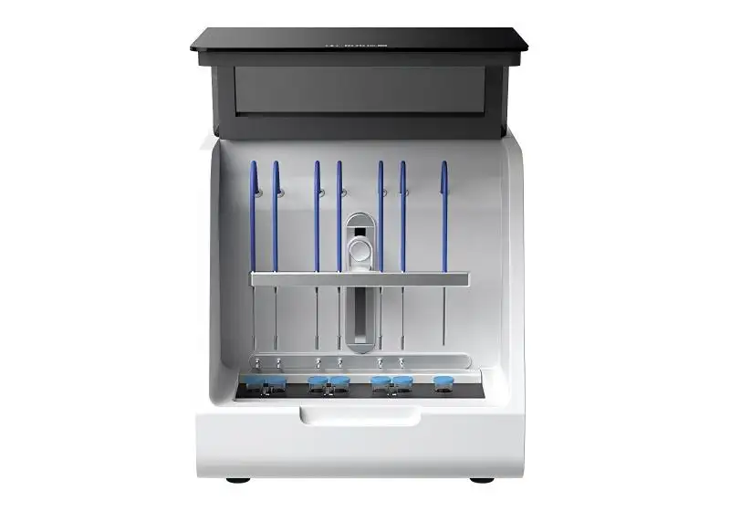
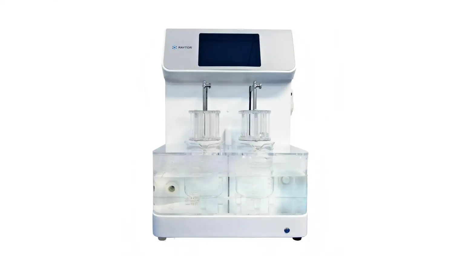
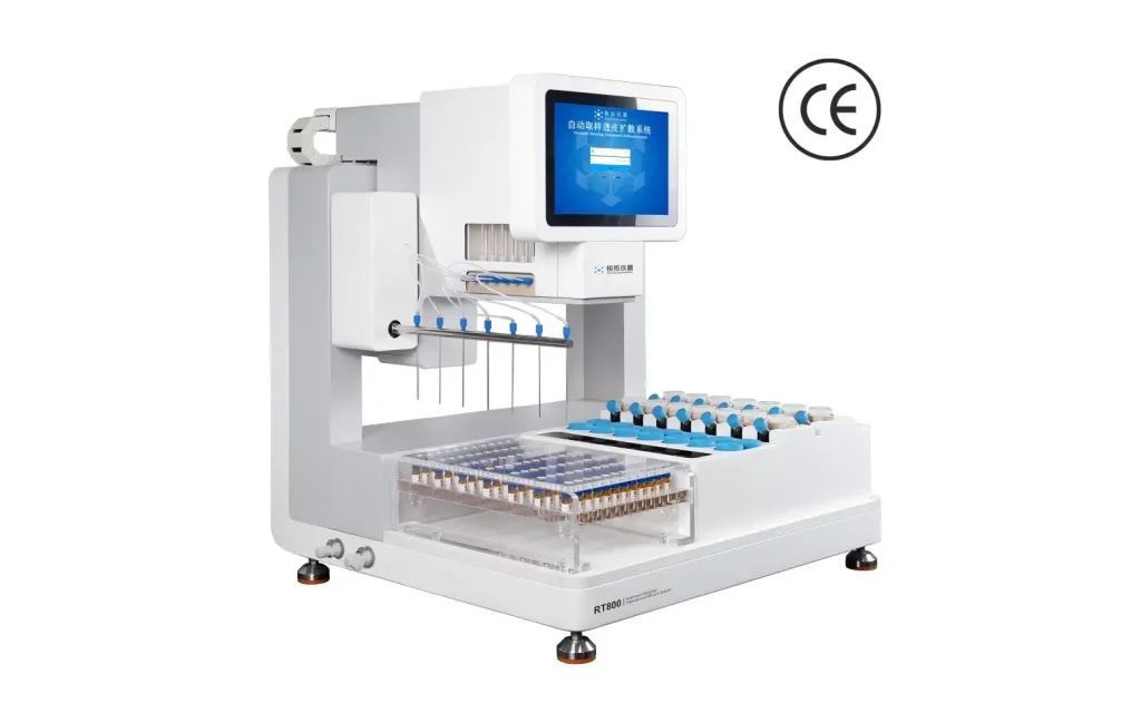
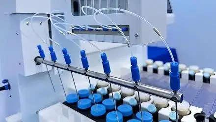
Raytor lessens this variability by automating critical functions of diffusion cell testing:
• Exact timing in sampling/refilling
• Automated sampling/refilling
• Short sampling paths reduce sample carryover and dead volume
• Diffusion cells allow inspection of bubbles and are easily loaded
• Filling of the receptor is easily confirmed by marks
• Cells can be operated independently for multiple comparisons
• Additional cell capacity allows a blank cell or control to be included
• Community standards of method control and adjust consistency of operation
• Better data recording and user/method controls allow improved traceability
• Automation eliminates most of the variation but does not replace required research and testing for product-specific applications.
When research has determined appropriate testing requirements, automating the testing protocols reduces the most significant variable of the testing process.
Special Considerations for Ophthalmic Suspensions
For complex ophthalmic suspensions, developers should confirm that:
• The membrane retains undissolved particles.
• Sampling measures the intended drug fraction.
• Particle dissolution can be distinguished from matrix release.
• The method detects relevant formulation changes.
• The selected Franz diffusion cell is appropriate for the product.
A conventional diffusion-cell method may not fully characterize every complex dispersed formulation. Alternative separation or release-testing strategies may be necessary when particle behavior dominates the result.
Does IVRT Predict Ophthalmic Bioavailability?
In Vitro Release Testing IVRT does not independently demonstrate ophthalmic bioavailability. Ocular exposure is also influenced by blinking, tear dilution, nasolacrimal drainage, corneal barriers, drug permeability, and formulation residence time.
IVRT results should therefore be interpreted with other product-quality, permeation, pharmacokinetic, or clinical evidence.
Final Words
As presented, specific IVRT methods will need to be developed for Ophthalmic ointments, gels, and suspensions. Consistent testing will depend on having an adequate formulation understanding, selecting an appropriate membrane and receptor medium, stabilizing the cell, and establishing a reproducible sampling protocol.
Raytor has developed automated systems for diffusion tests that help pharmaceutical laboratories eliminate the risk of residual volumes and improve the consistency and traceability of the IVRT testing. Raytor has the knowledge to help you decide whether your formulation and ophthalmic testing objectives would benefit from their systems.
FAQs
Q1. What types of formulations can Raytor systems support in IVRT studies?
Raytor systems can facilitate In Vitro Release Testing (IVRT) and IVPT for semi-solid and related formulations, such as ointments, gels, creams, patches, and some dispersed products. For other formulations, the method must first be validated.
Q2. Can Raytor equipment be used for ophthalmic ointment release testing?
Raytor systems can be used to provide a controlled environment for the release testing of ophthalmic ointments. It is the responsibility of the researcher to set up appropriate conditions for loading the samples, as well as to choose a membrane, receptor medium, sampling interval, and testing temperature.
Q3. Can Raytor systems test ophthalmic gels?
Raytor systems can test ophthalmic gels using methods that involve diffusion cells. To minimize variability, the system can be set to automate sampling and controlled agitation and change the medium accordingly.
Q4. Are Raytor systems suitable for ophthalmic suspensions?
Raytor systems, with some limitations, may be able to perform some studies on ophthalmic suspensions. However, issues related to the suspension, such as particle retention, sedimentation, dissolution rate, and the size of the membrane pores and sampling selectivity, should be considered.
Q5. How does synchronized sampling improve In Vitro Release Testing IVRT?
Since rapidly changing release rates require a sampling process that must be executed with extremely controlled timing, synchronized sampling is best for IVRT. The same can be said for closely spaced sampling where many diffusion cells must be sampled.